 Cardionerds: A Cardiology Podcast
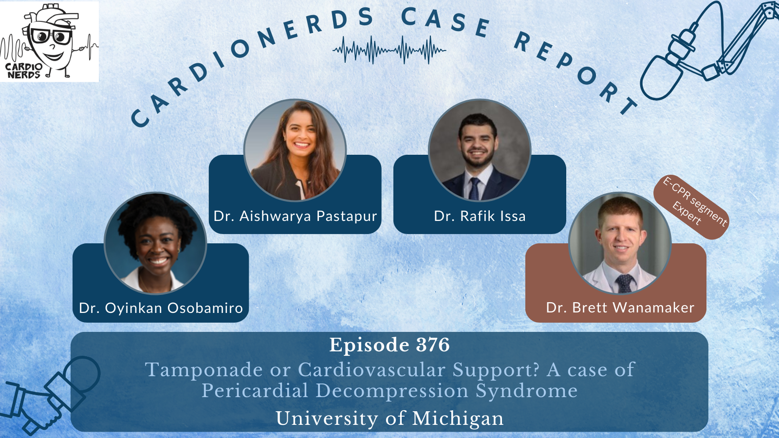
 376. Case Report: Tamponade or Cardiovascular Support? A case of Pericardial Decompression Syndrome – University of Michigan
CardioNerds cofounders, Dan Ambinder joins Drs. Aishwarya Pastapur, Oyinkansola Osobamiro, and Rafik Issa from the University of Michigan for drinks in Ann Arbor. They discuss the following case of pericardial decompression syndrome. Expert commentary is provided by Dr. Brett Wanamaker. Notes were drafted by Dr. Aishwarya Pastapur and Dr. Rafik Issa. The episode audio was engineered by CardioNerds Intern student Dr. Atefeh Ghorbanzadeh.
A woman in her 50s with a past medical history of stage IV lung cancer (with metastatic involvement of the liver, bone, and brain), previous saddle pulmonary emboli, pericardial effusion, and malignant pleural effusions presents with dyspnea. She was found to have a pericardial effusion with tamponade physiology relieved by pericardiocentesis. We discuss the management of cardiac tamponade, indications for pericardiocentesis, how to monitor for post-pericardiocentesis complications, and what to keep on your differential diagnosis for decompensation after pericardiocentesis. We discuss the epidemiology, pathophysiology, diagnosis, and management of pericardial decompression syndrome.
“To study the phenomena of disease without books is to sail an uncharted sea, while to study books without patients is not to go to sea at all.” – Sir William Osler. CardioNerds thank the patients and their loved ones whose stories teach us the Art of Medicine and support our Mission to Democratize Cardiovascular Medicine.
US Cardiology Review is now the official journal of CardioNerds! Submit your manuscript here.

Case Media – Pericardial Decompression Syndrome


Pearls – Pericardial Decompression Syndrome
- Diminished heart sounds, a low-voltage EKG with electrical alternans, elevated jugular venous pressure/pulsations (JVP), and the presence of pulses paradoxes are important findings that could suggest tamponade.
- McConnell sign is strongly concerning for right ventricular failure and pulmonary hypertension, potentially due to acute pulmonary embolism.
- Mechanical thrombectomy for pulmonary embolism is not feasible if the emboli are diffusely scattered without a central lesion to target.
- For patients who experience decompensation following pericardiocentesis, consider perforation, tamponade re-accumulation, or pericardial decompression syndrome (PDS).
- When possible, avoid draining more than 1L of pericardial fluid at once to minimize the risk of PDS.
Notes – Pericardial Decompression Syndrome
What is Pericardial Decompression Syndrome (PDS), and how does it present?
- Pericardial decompression syndrome is a rare, life-threatening syndrome occurring in about 5-10% of cases with paradoxical worsening of hemodynamics after pericardial drainage.
- The clinical presentation ranges from pulmonary edema to cardiogenic shock to death, occurring a few hours to days after a successful pericardiocentesis.
What is the underlying mechanism for PDS?
The pathophysiology behind PDS is debated, but there are three proposed mechanisms:
- Paradoxical Hemodynamic Derangement: After pericardiocentesis, venous return to the RV rapidly increases, resulting in RV expansion and potentially septal deviation towards the LV. Subsequently, the LV experiences decreased preload while still facing increased afterload as a compensatory response to obstructive shock, leading to decompensation.
- Myocardial Ischemia: Increased intrapericardial pressure may impair coronary perfusion, leading to myocardial ischemia. Upon pericardiocentesis, there is myocardial stunning with increased demand due to increased venous return and cardiac output
- Sympathetic Withdrawal: Withdrawal of sympathetic activation after drainage of pericardial fluid can trigger cardiovascular collapse
What are the risk factors for developing PDS, and how can we mitigate those risks for prevention?
- Generally, patients with long-standing pericardial effusion with chronic compression of the heart, such as those with malignant pericardial effusions, are more vulnerable to developing PDS after pericardiocentesis.
- Additionally, rapid fluid removal increases the risk. In terms of prevention, removing fluid to normalize CVP and MAP and letting the rest of the fluid drain slowly may mitigate the risk.
How do we manage a patient with PDS?
- The management of PDS is supportive, focusing on addressing hemodynamic and respiratory derangements.
- The underlying pathophysiology should resolve in 24-48 hours.
What is the prevalence and prognosis of PDS?
- PDS affects 5-10% of pericardiocentesis procedures, although the exact frequency is difficult to ascertain.
- It is a self-resolving process as the heart re-adapts to the new hemodynamics.
- However, during the episode of PDS, mortality can be as high as 30% per some studies.
References – Pericardial Decompression Syndrome
- Schnur M. Understanding Pulsus Paradoxus. Accessed February 27, 2024. https://nursingcenter.com/ncblog/august-2021/understanding-pulsus-paradoxus
- Carlini’ ’Caterina Chiara De, Maggiolini’ ’Stefano. Pericardiocentesis in cardiac tamponade: indications and practical aspects. Accessed February 27, 2024. https://www.escardio.org/Journals/E-Journal-of-Cardiology-Practice/Volume-15/Pericardiocentesis-in-cardiac-tamponade-indications-and-practical-aspects
- Angouras DC, Dosios T. Pericardial Decompression Syndrome: A Term for a Well-Defined but Rather Underreported Complication of Pericardial Drainage. The Annals of Thoracic Surgery. 2010;89(5):1702-1703. doi:10.1016/j.athoracsur.2009.11.073
- Imazio M. Pericardial decompression syndrome: A rare but potentially fatal complication of pericardial drainage to be recognized and prevented. European Heart Journal Acute Cardiovascular Care. 2015;4(2):121-123. doi:10.1177/2048872614557771
- Prabhakar Y, Goyal A, Khalid N, et al. Pericardial decompression syndrome: A comprehensive review. World Journal of Cardiology. 2019;11(12):282-291. doi:10.4330/wjc.v11.i12.282
- Sobieski C, Herner M, Goyal N, et al. Pericardial Decompression Syndrome After Drainage of Chronic Pericardial Effusions. JACC: Case Reports. 2022;4(22):1515-1521. doi:10.1016/j.jaccas.2022.08.023
- Chhabra L. Pericardial Decompression Syndrome. American College of Cardiology. Accessed February 27, 2024. https://www.acc.org/Latest-in-Cardiology/Articles/2020/04/13/09/05/http%3a%2f%2fwww.acc.org%2fLatest-in-Cardiology%2fArticles%2f2020%2f04%2f13%2f09%2f05%2fPericardial-Decompression-Syndrome
- Pradhan R, Okabe T, Yoshida K, Angouras DC, DeCaro MV, Marhefka GD. Patient characteristics and predictors of mortality associated with pericardial decompression syndrome: a comprehensive analysis of published cases. European Heart Journal Acute Cardiovascular Care. 2015;4(2):113-120. doi:10.1177/2048872614547975
- Amro A, Mansoor K, Amro M, et al. A Comprehensive Systemic Literature Review of Pericardial Decompression Syndrome: Often Unrecognized and Potentially Fatal Syndrome. Current Cardiology Reviews. 17(1):101-110.
