Cardionerds: A Cardiology Podcast
 87. Case Report: Giant Coronary Aneurysm Presenting with Heart Failure – University of Hawaii
Aloha! CardioNerds (Amit Goyal & Karan Desai) join University of Hawaii cardiology fellows (Isaac Mizrahi, Nath Limpruttidham, Nishant Trivedi, and Shana Greif) for some shaved iced on the Big Island’s north shore! They discuss a fascinating case of a patient presenting with decompensated heart failure found to have a giant coronary aneurysm. Program director Dr. Dipanjan Banerjee provides the E-CPR as well as a message for applicants. Episode notes were developed by Johns Hopkins internal medicine resident Tommy Das with mentorship from University of Maryland cardiology fellow Karan Desai.
Jump to: Patient summary – Case media – Case teaching – References

CardioNerds Case Reports Page
CardioNerds Episode Page
CardioNerds Academy
Subscribe to our newsletter- The Heartbeat
Support our educational mission by becoming a Patron!
Cardiology Programs Twitter Group created by Dr. Nosheen Reza
Patient Summary
A man in his early 60s with history of hypertension, peripheral arterial disease, atrial fibrillation, and AAA s/p repair presented with subacute fatigue, palpitations, shortness of breath, and lower extremity edema. On exam he was warm and well perfused, though hypotensive, tachycardic with an irregular rhythm, and had an elevated JVP. ECG showed AF with RVR without evidence of acute MI, and troponin was negative. TTE revealed a reduced LVEF and WMA in the inferolateral walls with akinesis of the basal mid septum; additionally, two large extracardiac structures were noted, one with heterogenous echotexture in the AV groove, and a second with an echolucent interior adjacent to the RA.
The patient underwent coronary angiography, showing a dilated and calcified proximal LAD with high grade stenosis adjacent to the first septal perforator, a ectatic LCX that supplied left to right collaterals, and a giant RCA aneurysm with TIMI 0 flow distally. CCTA confirmed these findings, showing thrombosed aneurysms of the LAD, LCX, and RCA. Interventional cardiology and cardiac surgery both evaluated the patient’s case, and determined that he was not a candidate for intervention. He was ultimately diuresed to euvolemia with significant improvement in symptoms, and plans to follow-up as an outpatient for heart transplant evaluation.
Case Media
-

A -

B -

C -

D
A. CXR
B. ECG: atrial fibrillation with RVR, left axis deviation, poor r wave progression
C. Wide complex tachycardia
D. CT chest demonstrating giant aneurysm
Episode Schematics & Teaching
The CardioNerds 5! – 5 major takeaways from the #CNCR case
1) This case featured a patient with a giant coronary aneurysm – how are coronary artery aneurysms defined and classified?
- Coronary artery aneurysms (CAA) are defined as a focal dilation of a coronary segment at least 1.5x the adjacent normal segment. Contrast this with coronary artery ectasia, which refers to a diffuse, as opposed to focal, coronary dilation.
- CAA morphology can be classified as either saccular (transverse > longitudinal diameter) or fusiform (transverse < longitudinal diameter).
- Giant CAA’s are >20mm in diameter.
- Aortocoronary saphenous vein graft aneurysms have distinct characteristics and natural history compared to native coronary aneurysms. These aneurysms tend to present late (e.g., > 10 years following CABG) and tend to be larger than native CAA.
- IVUS can help differentiate between a true aneurysm with preserved integrity of all 3 vessel layers (intima, media, and adventitia) and a pseudoaneurysm with loss of wall integrity and damage to the adventitia.
2) Now that we have the language to define and classify coronary artery aneurysms, what are some causes these lesions?
- Atherosclerosis: lipid deposition, focal calcification, and fibrosis can weaken the vessel wall and predispose to subsequent coronary artery dilation. Up to 50% of CAAs are linked to arteriosclerosis.
- Autoimmune/inflammatory processes: Lupus and systemic vasculitis, such as Kawasaki’s disease and Takayasu arteritis, can all lead to CAAs. Vasculitic CAAs usually affect more than one artery.
- Connective Tissue Disease: Marfan’s syndrome and Ehlers-Danlos disease, for instance, are characterized by deficiencies in vessel wall integrity, leading to CAAs.
- Dynamic Wall Stress: Episodic hypertension and vasoconstriction, such as that seen in frequent cocaine use, can lead to wall stress, endothelial damage, and coronary artery aneurysms.
- Direct Vessel Wall Injury: Intracoronary interventions, such as angioplasty, stent delivery, and brachytherapy, can cause shear wall stress that leads to CAAs.
- Infectious Causes: Direct vessel wall invasion or immune complex deposition can be seen in bacterial, mycobacterial, fungal, and syphilitic infections. Septic emboli from infectious endocarditis can also lead to mycotic coronary aneurysms.
- Genetic susceptibility: Certain HLA class II genotypes are susceptible to CAAs. This may be the underlying pathology in certain idiopathic and congenital CAAs.
3) How do coronary artery aneurysms clinically present?
- Most CAAs are asymptomatic, and are found incidentally on coronary angiography or CCTA.
- Concomitant obstructive arteriosclerosis can cause angina or plaque rupture, and thrombosis in the aneurysm lumen can lead to distal embolization and myocardial infarction.
- Massive enlargement of CAAs and saphenous vein graft aneurysms can compress adjacent structures.
- CAA rupture is rare, though can cause cardiac tamponade.
- Stress-induced myocardial ischemia can also occur due to microvascular dysfunction
4) How do we diagnosis and assess CAAs?
- Most CAAs are evaluated via coronary angiography, though a complete angiographic evaluation can be complicated by delayed antegrade contrast filling, segmental back flow, and contrast stasis. In giant aneurysms, a forceful and prolonged contrast injection is needed to avoid misinterpreting slow aneurysmal filling as thrombosis.
- IVUS can help differentiate between true aneurysms, pseudoaneurysms, and coronary segments with aneurysmal appearance due to plaque rupture or stenosis. Furthermore, IVUS can assist in sizing aneurysm and planning for potential PCI.
- Coronary CTA noninvasively allows for a more accurate assessment of aneurysm size and degree of thrombus than angiography. CCTA is particularly useful in patients with giant CAA as it can provide an understanding of mechanical complications of these aneurysms.
5) How are coronary artery aneurysms managed?
- Notably, there is a lack of randomized and large-scale trial data to guide the treatment of CAAs; most recommendations are made on the basis of small case series and expert opinion.
- Medical Management: Given the association between CAA and arteriosclerosis, risk factor modification should be emphasized. The role of antiplatelet and anticoagulant agents is an area of ongoing debate, though there may be benefit in patients with multivessel ectasia. Furthermore, the context in which the patient presents (e.g., incidental finding vs. acute coronary syndrome) will guide the antiplatelet and/or anticoagulant strategy.
- Invasive (Percutaneous) Management: PCI of an aneurysmal vessel in the setting of acute MI is associated with lower rates of procedural success, and higher rates of distal embolization and no-reflow phenomenon. Additionally, these patients have higher rates of stent thrombosis and mortality. Given the higher thrombus burden in aneurysmal arteries, thrombectomy may be helpful in aiding PCI. Some case studies have additionally utilized intracoronary thrombolytics. Another strategy is a stent-assisted coil embolization technique in cases where covered stent placement is not possible due to tortuosity, calcification, or risk of side branch compromise. To date, there haven’t been covered stents specifically designed for CAAs, but stents have been used off-label.
- Surgical Management: The most common operative strategy is to open the aneurysm, suture the afferent and efferent vessels, and finish with bypass grafting if possible. Other operative strategies include aneurysm ligation, resection, or marsupialization with interposition graft.
References
- Thibodeau, J. T., & Drazner, M. H. (2018). The Role of the Clinical Examination in Patients With Heart Failure. JACC. Heart failure, 6(7), 543–551.
- Abou Sherif, S., Ozden Tok, O., Taşköylü, Ö., et al. (2017). Coronary Artery Aneurysms: A Review of the Epidemiology, Pathophysiology, Diagnosis, and Treatment. Frontiers in cardiovascular medicine, 4, 24.
- Kawsara, A., Núñez Gil, I. J., Alqahtani, F., et al. (2018). Management of Coronary Artery Aneurysms. JACC. Cardiovascular interventions, 11(13), 1211–1223.
- Newburger, J. W., Takahashi, M., & Burns, J. C. (2016). Kawasaki Disease. Journal of the American College of Cardiology, 67(14), 1738–1749.
- McCrindle, B. W., Rowley, A. H., Newburger, J. W., et al. (2017). Diagnosis, Treatment, and Long-Term Management of Kawasaki Disease: A Scientific Statement for Health Professionals From the American Heart Association. Circulation, 135(17), e927–e999.
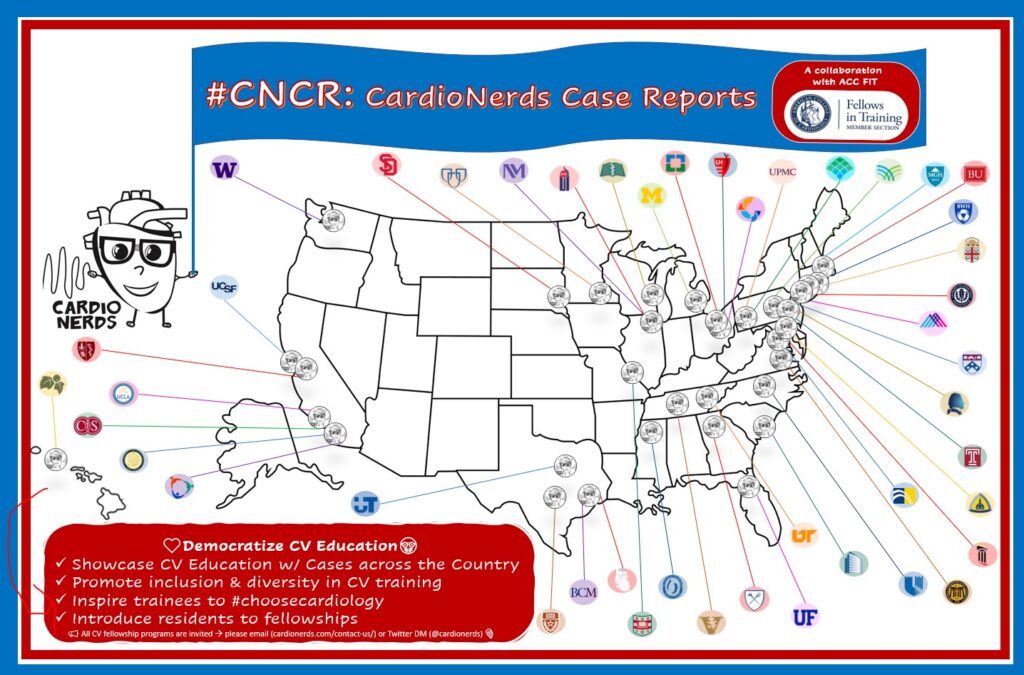
The CardioNerds Cardiology Case Reports series shines light on the hidden curriculum of medical storytelling. We learn together while discussing fascinating cases in this fun, engaging, and educational format. Each episode ends with an “Expert CardioNerd Perspectives & Review” (E-CPR) for a nuanced teaching from a content expert. We truly believe that hearing about a patient is the singular theme that unifies everyone at every level, from the student to the professor emeritus.
We are teaming up with the ACC FIT Section to use the #CNCR episodes to showcase CV education across the country in the era of virtual recruitment. As part of the recruitment series, each episode features fellows from a given program discussing and teaching about an interesting case as well as sharing what makes their hearts flutter about their fellowship training. The case discussion is followed by both an E-CPR segment and a message from the program director.
CardioNerds Case Reports: Recruitment Edition Series Production Team
-

Bibin Varghese, MD -

Rick Ferraro, MD -
Tommy Das, MD -

Eunice Dugan, MD -

Evelyn Song, MD -

Colin Blumenthal, MD -
Karan Desai, MD -

Amit Goyal, MD -

Daniel Ambinder, MD