This episode delves into pediatric c-spine injuries focusing on the question of who needs imaging?

 Core EM - Emergency Medicine Podcast
 Episode 30.0 – Pediatric C-spine Injuries
Jan 18, 2016
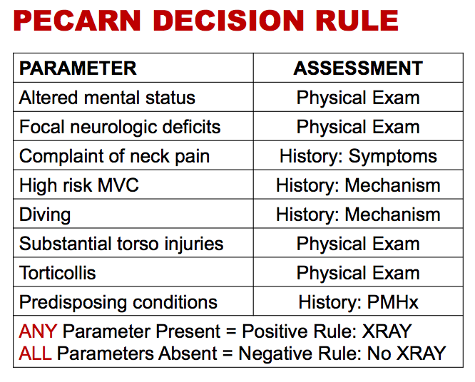
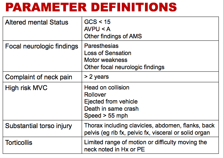
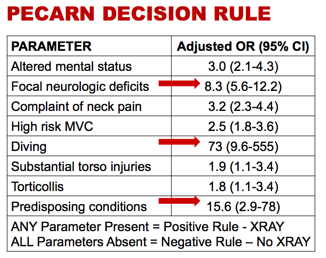
Explore the intricate world of pediatric cervical spine injuries and the critical question of imaging necessity for young patients. The hosts tackle the unique anatomical challenges in diagnosing these injuries and the complications that arise. A mnemonic for imaging guidelines is introduced, along with insights into the PCARN algorithm. Practical tips for handling pediatric patients, especially in bulky winter clothing, provide an entertaining twist as they combine serious medical insights with humorous anecdotes.
AI Snips
Chapters
Transcript
Episode notes
Avoid Cutting Down Coats
- Do not cut down coats with trauma shears during emergency care.
- Removing these coats improperly creates a mess with feathers and complicates patient management.
Pediatric Anatomy Changes Injury Patterns
- Pediatric c-spine injuries differ from adults due to anatomical factors like weak neck muscles and ligament laxity.
- These differences also make x-ray interpretation more challenging due to variations like incomplete physis closure.
Fractures Concentrate at C1-C2
- Most pediatric cervical spine fractures occur in the C1 and C2 vertebrae.
- Lower cervical spine injuries are very rare in children, supporting limited imaging approaches focused on the upper c-spine.