Dr. Vishwajit Hegde, a second-year Pulmonary and Critical Care fellow, shares a captivating case of a 26-year-old with chronic cough and pulmonary nodules. Dr. Sahil Pandya, an Associate Professor and program director, dives into diagnostic strategies, emphasizing a Bayesian approach to avoid premature conclusions. They discuss interpreting intricate imaging patterns and the decision-making process for bronchoscopic tissue sampling. The big reveal? A diagnosis of miliary lung adenocarcinoma leading to a targeted therapy breakthrough. Insights into KU's fellowship culture shine as both guests emphasize mentorship and trainee education.
00:00
forum Ask episode
web_stories AI Snips
view_agenda Chapters
auto_awesome Transcript
info_circle Episode notes
question_answer ANECDOTE
Young Immigrant Treated Empirically For TB
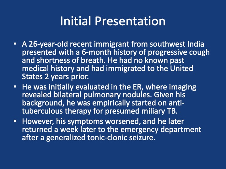
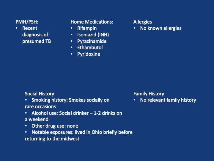
A 26-year-old immigrant from Southwest India had six months of progressive cough and nodular lung disease and was empirically started on RIPE for presumed miliary TB.
He returned two weeks later with a generalized tonic-clonic seizure, prompting reevaluation and further workup.
volunteer_activism ADVICE
Confirm TB Before Relying On Empiric Therapy
Always confirm tuberculosis when possible with microbiology or pathology and obtain resistance testing rather than relying solely on empiric RIPE.
Use a Bayesian approach: combine pre-test probability with targeted tests before committing to long treatments.
insights INSIGHT
Interval Change Upweights Infection Or Aggressive Malignancy
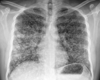
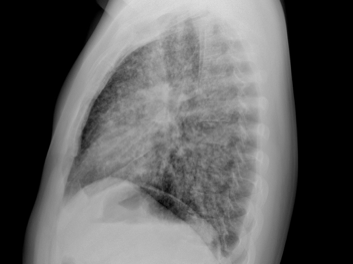
Interval CT showed diffuse miliary nodules developing central cavitation, confluent areas, and septal thickening which raised concern for infection or aggressive malignancy.
New cavitation and confluence on interval imaging should increase suspicion for infection or aggressive cancer.
Get the Snipd Podcast app to discover more snips from this episode
After a brief hiatus, we are excited to be back today with another Fellows’ Case Files! Today we’re virtually visiting the University of Kansas Medical Center (KUMC) to hear about a fascinating pulmonary presentation. There are some fantastic case images and key learning points. Take a listen and see if you can make the diagnosis along with us. As always, let us know your thoughts and definitely reach out if you have an interesting case you’d like to share.
Meet Our Guests
Dr. Vishwajit Hegde completed his internal medicine residency at University of Kansas Medical Center where he stayed for fellowship and is currently a second year Pulmonary and Critical Care medicine fellow.
Dr. Sahil Pandya is an Associate Professor of Medicine and Program Director of the PCCM Fellowship at KUMC.
Case Presentation
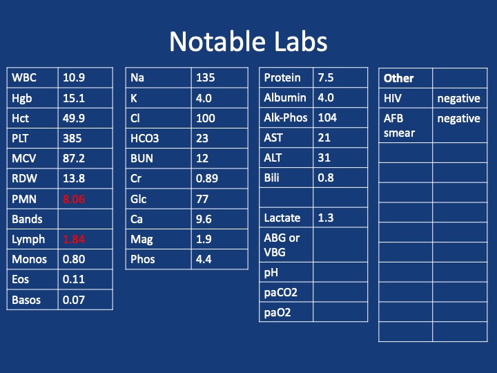
Imaging
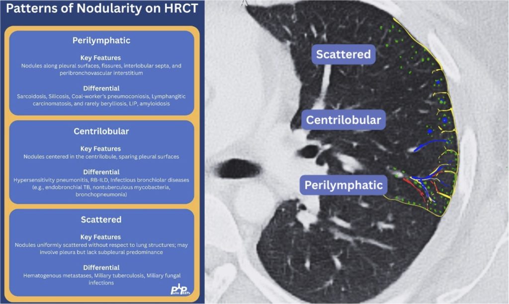
Infographic
Key Learning Points
1) Initial frame & diagnostic mindset
Young (26), subacute → chronic dyspnea/cough with diffuse pulmonary nodules; avoid premature closure on TB.
Use a Bayesian approach: combine pre-test probability (epidemiology, exposures, tempo) with targeted tests to decide next steps.
Always confirm TB when possible (micro/path + resistance testing); empiric RIPE may be reasonable but shouldn’t replace tissue when stakes are high.
2) Imaging pearls—nodular pattern recognition
Ask three things: craniocaudal distribution, symmetry, central vs peripheral.
Partner with neuroradiology for pattern nuances; treat seizures but keep searching for the unifying diagnosis.
4) Lab/serology strategy
Broad infectious workup (AFB × multiple, fungal serologies), HIV and basic immune screen.
Negative/indeterminate tests don’t end the search—revisit history (e.g., Ohio travel → histo/blasto risk).
5) “Tissue is the issue”—choosing the procedure
For diffuse nodules with mediastinal adenopathy and stable patient: EBUS-TBNA + BAL, consider transbronchial or cryobiopsy.
Cryobiopsy pros: larger, less crush artifact, better for molecular testing; cons: ↑ bleeding/pneumothorax vs forceps.
VATS still best for certain ILD questions or if less invasive routes are non-diagnostic—but weigh patient preference and stage/likelihood of yield.
6) ROSE (rapid on-site evaluation) in bronchoscopy
Confirms adequacy in real time, steers you away from necrotic zones, helps decide when you’ve got enough for molecular studies, and when to pivot sites—reduces anesthesia time and repeat procedures.
7) Final diagnosis & management
Path: TTF-1+, CK7+, napsin A → pulmonary adenocarcinoma with a fusion driver.
Therapy: Targeted TKI (crizotinib) → dramatic radiographic response of miliary lung disease and CNS lesions.
Teaching point: even “miliary TB-like” lungs + CNS lesions in a 20-something can be driver-positive lung cancer—don’t let age or pattern blind you.
References and Further Reading
Desai, S., Devaraj, A., Lynch, D., & Sverzellati, N. (2020). Webb, Müller and Naidich’s high-resolution CT of the lung (6th ed.). Lippincott Williams & Wilkins.
Rajeswaran, G., Becker, J. L., Michailidis, C., Pozniak, A. L., & Padley, S. P. G. (2006). The radiology of IRIS (immune reconstitution inflammatory syndrome) in patients with mycobacterial tuberculosis and HIV co-infection: appearances in 11 patients. Clinical radiology, 61(10), 833-843
Poletti, V., Ravaglia, C., & Tomassetti, S. (2016). Transbronchial cryobiopsy in diffuse parenchymal lung diseases. Current opinion in pulmonary medicine, 22(3), 289-296.
Norman, G. R., Monteiro, S. D., Sherbino, J., Ilgen, J. S., Schmidt, H. G., & Mamede, S. (2017). The causes of errors in clinical reasoning: cognitive biases, knowledge deficits, and dual process thinking. Academic Medicine, 92(1), 23-30.

 PulmPEEPs