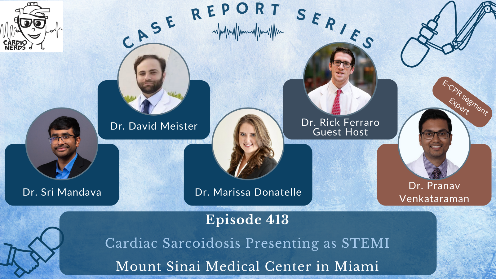
413. Case Report: Cardiac Sarcoidosis Presenting as STEMI – Mount Sinai Medical Center in Miami
Cardionerds: A Cardiology Podcast
Case Study of Acute Chest Pain in a 57-Year-Old Male
This chapter presents a detailed case study of a 57-year-old man experiencing severe chest pain at night, leading to an emergency room visit. The speakers analyze the patient's medical history and lifestyle factors to explore the implications for cardiovascular health.
CardioNerds (Dr. Rick Ferraro and Dr. Dan Ambinder) join Dr. Sri Mandava, Dr. David Meister, and Dr. Marissa Donatelle from the Columbia University Division of Cardiology at Mount Sinai Medical Center in Miami. Expert commentary is provided by Dr. Pranav Venkataraman. They discuss the following case involving a patient with cardiac sarcoidosis presenting as STEMI.

A 57-year-old man with a history of hyperlipidemia presented with sudden onset chest pain. On admission, he was vitally stable with a normal cardiorespiratory exam but appeared in acute distress and was diffusely diaphoretic. His ECG revealed sinus rhythm, a right bundle branch block (RBBB), and ST elevation in the inferior-posterior leads. He was promptly taken for emergent cardiac catheterization, which identified a complete thrombotic occlusion of the mid-left circumflex artery (LCX) and large obtuse marginal (OM) branch, with no underlying coronary atherosclerotic disease. Aspiration thrombectomy and percutaneous coronary intervention (PCI) were performed, with one drug-eluting stent placed. An echocardiogram showed a left ventricular ejection fraction (EF) of 31%, hypokinesis of the inferior, lateral, and apical regions, and an apical left ventricular thrombus. The patient was started on triple therapy. A hypercoagulable workup was negative. A cardiac MRI was obtained to further evaluate non-ischemic cardiomyopathy. In conjunction with a subsequent CT chest, the results raised suspicion for cardiac sarcoidosis with systemic involvement. In view of a reduced EF and significant late-gadolinium enhancement, electrophysiology was consulted to evaluate for ICD candidacy. A decision was made to delay ICD implantation until a definitive diagnosis of cardiac sarcoidosis could be established by tissue biopsy. The patient was started on HF-GDMT and discharged with a LifeVest. Close outpatient follow-up with cardiology and electrophysiology was arranged.
“To study the phenomena of disease without books is to sail an uncharted sea, while to study books without patients is not to go to sea at all.” – Sir William Osler. CardioNerds thank the patients and their loved ones whose stories teach us the Art of Medicine and support our Mission to Democratize Cardiovascular Medicine.
US Cardiology Review is now the official journal of CardioNerds! Submit your manuscript here.
Pearls – Cardiac Sarcoidosis Presenting as STEMI
- Cardiac sarcoidosis can present with a variety of symptoms, including arrhythmias, heart block, heart failure, or sudden cardiac death. Symptoms can be subtle or mimic other cardiac conditions.
- Conduction abnormalities, particularly AV block or ventricular arrhythmias, are common and may be the initial indication of cardiac involvement with sarcoidosis.
- The additive value of Echocardiography, FDG-PET, and cardiac MR is indispensable in the diagnostic workup of suspected cardiac sarcoidosis.
- Specific role of MRI/PET: Both cardiac MRI and FDG-PET provide a complementary role in the diagnosis of cardiac sarcoidosis. Cardiac MRI is an effective diagnostic screening tool with fairly high sensitivity but is limited by its inability to decipher inflammatory (“active” disease) versus fibrotic myocardium. FDG-PT helps to make this discrimination, refine the diagnosis, and guide clinical management. Ultimately, these studies are most useful when interpreted in the context of other clinical information.
- Primary prevention of sudden cardiac death in cardiac sarcoidosis focuses on risk stratification, with ICD placement for high-risk patients. For patients awaiting definitive diagnosis, a LifeVest may be used as a temporary measure to protect from sudden arrhythmic events until an ICD is placed.
Notes – Cardiac Sarcoidosis Presenting as STEMI
1. Is STEMI always a result of coronary artery disease?
By definition, a STEMI is an acute S-T segment elevation myocardial infarction. This occurs when there is occlusion of a major coronary artery, which results in transmural ischemia and damage, resulting in electrical changes seen on the ECG. The most common cause of coronary artery occlusion is coronary artery disease (CAD) from plaque rupture and thrombus formation; however, many other causes of coronary artery occlusion are not related to CAD. These include vasospasm (isolated and recurrent), in-situ thrombotic occlusion, spontaneous coronary artery dissection, and supply-demand mismatch, such as in the setting of severe anemia. DDx includes other causes of injury current, such as myocarditis. It is important to keep these other differentials in mind while preparing for coronary angiography, as it may help guide intra-catheterization and post-catheterization management.
2. What are the most common causes of LV thrombus?
When considering the causes of thrombus formation, think of Virchow’s triad. As with any other location, thrombus formation in the LV may be caused by injury/inflammation, systemic thrombophilia, and stasis.
- Acute myocardial infarction (especially anterior MI) – damaged myocardium and impaired LV function lead to blood stasis and thrombus formation.
- Heart failure with reduced ejection fraction (HFrEF) – severely impaired contractility increases the risk of thrombus development
- Non-Ischemic cardiomyopathies – dilated or hypertrophic cardiomyopathies may cause abnormal blood flow, promoting thrombus formation.
- Arrhythmias – although more associated with atrial thrombus, atrial fibrillation can also contribute to LVT in cases of significant LV dysfunction. Ventricular arrhythmias can also cause LV thrombus.
- Hypercoagulable conditions – Conditions such as antiphospholipid antibody syndrome, inherited thrombophilias, malignancy-associated hypercoagulability, polycythemia vera, hyperhomocysteinemia, nephrotic syndrome or systemic lupus erythematous may predispose to LV thrombus formation
- Inflammatory conditions – conditions like myocarditis or cardiac sarcoidosis can lead to inflammation along with focal stasis from aneurysmal changes, contributing to thrombus formation
3. What is the clinical presentation of cardiac sarcoidosis?
- Chest pain: can arise from several mechanisms such as myocardial inflammation, pericarditis, coronary artery involvement, or arrhythmias.
- Heart Failure: symptoms such as dyspnea, fatigue, and peripheral edema may result from left ventricular dysfunction or restrictive cardiomyopathy.
- Arrhythmias: palpitations, dizziness or syncope may occur due to ventricular tachycardia or ventricular fibrillation.
- Conduction abnormalities: Heart block, especially complete AV block, is a common early manifestation. Some studies have found that AV block is the presenting symptom in more than 40% of patients with cardiac sarcoidosis.
- Sudden cardiac death (SCD): sudden death can occur due to ventricular arrhythmias or severe heart block.
- Asymptomatic: in some cases, cardiac sarcoidosis is discovered incidentally during imaging or evaluation for systemic sarcoidosis.
4. What are the key imaging modalities used in the diagnosis of cardiac sarcoidosis?
Echocardiography, FDG-PET, and cardiac MRI are the key imaging modalities used to diagnose cardiac sarcoidosis. The echocardiogram is often normal in clinically silent disease, but several key features may be seen in clinically active disease. The most specific findings are basal interventricular thinning and LV aneurysm. Other less specific findings include increased LV wall thickness, LV/RV diastolic and/or systolic dysfunction, and wall motion abnormalities (non-coronary distribution). Strain imaging is promising for use in earlier stages of disease, but this is not well established yet. FDG-PET is crucial in the initial diagnosis of cardiac sarcoidosis, allowing active inflammatory disease to be detected. There is no pathognomonic PET finding; however, focal or focal-on-diffuse FDG uptake patterns are highly suggestive of active disease. It should be noted that FDG-PET is also useful in guiding treatment or response to immunosuppressive therapy, as it can track the degree of inflammation over time. The role of cardiac MRI is discussed below.
5. What is the specific role of cardiac MRI in the diagnosis of cardiac sarcoidosis?
This depends on the specific clinical setting. A patient with established extra-cardiac sarcoidosis but asymptomatic from a cardiac standpoint should be appropriately screened for cardiac involvement by clinical history, ECG, echocardiography, and cardiac monitoring (e.g. Holter monitor, etc). If any of the aforementioned “screening” tests are abnormal, a cardiac MRI is then indicated to assess for evidence of cardiac sarcoidosis. More specifically, cardiac MRI detects inflammation and edema at earlier stages of disease and scar tissue at later stages. The classical finding specific for cardiac sarcoidosis is patchy late gadolinium enhancement, with a predilection for the basal septum and basal inferolateral wall. The enhancement is either subepicardial or mid-wall and rarely transmural. It should be noted that once cardiac sarcoidosis is diagnosed, FDG-PET imaging should be utilized in conjunction with, or complementary to MRI, to assess for “active sarcoid” (i.e. myocardial inflammation).
On the other hand, a patient with no known extracardiac sarcoidosis but with suggestive cardiac findings should have a cardiac MRI to assess for typical features as mentioned above, in addition to assessment for non-cardiac involvement.
It should be noted that cardiac MRI can also provide significant prognostic information. The presence of LGE portends a worse prognosis due to increased CV death and ventricular arrhythmias. It should also be noted that LGE does not discriminate between active inflammation and fibrosis. Tissue characterization with T1 and T2 mapping techniques or PET imaging, as described above, can be more useful in this sense.
References
1.) Cheng RK, Kittleson MM, Beavers CJ, et al. Diagnosis and management of cardiac sarcoidosis: a scientific statement from the American Heart Association. Circulation. 2024;149.
2.) Lehtonen J, Uusitalo V, Pöyhönen P, Mäyränpää MI, Kupari M. Cardiac sarcoidosis: phenotypes, diagnosis, treatment, and prognosis. European Heart Journal. 2023;44:1495–1510.
3.) Kouranos V, Sharma R. Cardiac sarcoidosis: state-of-the-art review. Heart. 2021;107:1591–1599.
4.) Birnie DH, Nery PB, Ha AC, Beanlands RSB. Cardiac sarcoidosis. Journal of the American College of Cardiology. 2016;68:411–421.


