
79. Case Report: Recurrent Troponin Elevation – University of Washington
Cardionerds: A Cardiology Podcast
Introduction
Explore a captivating case study presented by fellows from the University of Washington Cardiology Fellowship Program, aiming to democratize cardiovascular education and foster diversity in the field.
CardioNerds (Amit Goyal & Daniel Ambinder) join University of Washington cardiology fellows (Shannon McConnaughey, Betty Ashinne and Andrew Perry – host of the AP Cardiology podcast) for some tacos and beer at the water and discuss a puzzling case of recurrent troponin elevation. Dr. Kelly Branch provides the E-CPR and program director, Dr. Rosario Freeman, provides a message for applicants. Episode notes were developed by Johns Hopkins internal medicine resident Eunice Dugan with mentorship from University of Maryland cardiology fellow Karan Desai.
Jump to: Patient summary – Case media – Case teaching – References

CardioNerds Case Reports Page
CardioNerds Episode Page
CardioNerds Academy
Subscribe to our newsletter- The Heartbeat
Support our educational mission by becoming a Patron!
Cardiology Programs Twitter Group created by Dr. Nosheen Reza
Patient Summary
A female is her late 50s with past medical history of alcohol use disorder and intravenous drug use complicated by hepatitis C presented with generalized weakness and was found to have lower extremity fractures. Cardiology was consulted for pre-operative management. When obtaining a cardiac history, she reported rare, intermittent, and non-anginal chest pain. She had no current chest pain or dyspnea. On chart review, she had multiple presentations over the past 3 years for various complaints – some were chest pain – during which she was found to have elevated troponin I values. Most of the elevations were in the 0.5 to 1 ng/mL range, but one time, her peak troponin was 32 ng/mL. At one of those presentations, she had a TTE that showed septal wall motion abnormalities. She underwent coronary angiography on two occasions, which both showed proximal and mid-LAD calcifications, but no significant stenosis and she was managed medically.
At the current presentation, her vitals were within normal limits and her exam did not show any cardiopulmonary abnormalities. Labs were notable for troponin I of 10 ng/mL but CK-MB was normal. Interestingly, alkaline phosphatase was 3 times the upper limit of normal, and rheumatoid factor was 1156 IU/mL compared to 70 previously (normal range 0-20 IU/mL). EKG was without ischemic changes. TTE showed no wall motion abnormalities, normal LVEF, and no significant valvular disease. Cardiac catherization was deferred, and the patient was not interested in cardiac MRI. Due to an incongruent troponin elevation with the clinical presentation, there was concern for interference with the troponin assay due to the elevated rheumatoid factor and alkaline phosphatase levels. Upon further investigation, dilution of the troponin yielded results lower than factor of dilution or undetectable results. Mayo testing of the sample showed troponin-T of 0.024 ng/mL (normal < 0.01 ng/mL), and troponin I of 0.02 ng/mL (normal < 0.04 ng/mL). Although there were still some aspects of her previous presentations including rising troponin pattern and previous wall motion abnormalities that are unexplained at this time, her troponin elevation at this presentation was attributed to assay interference.
Case Media
-

A -

B -

C
A. ECG from May
B. Coronary angiography form May
C. ECG August (same year)
Episode Schematics & Teaching
Coming soon!
The CardioNerds 5! – 5 major takeaways from the #CNCR case
- What is the universal definition of myocardial infarction?
· As per the current 4th universal definition, myocardial infarction is defined as an elevation in cardiac troponin (cTn) above the 99th percentile in high sensitivity assays in the setting of acute myocardial ischemia. It is considered acute if there is a characteristic rise/fall pattern in cTn.
· A type 1 event is a spontaneous MI related to ischemia as a result of a primary coronary event (e.g., plaque erosion or rupture, dissection). The definition requires the above biomarker threshold with at least one of the following as evidence of ischemia: symptoms of MI, new ischemic EGG changes including development of pathological Q waves, new imaging evidence of loss of viable myocardium, or identification of thrombosis by intracoronary imaging. Unstable angina (UA) occurs in patients with symptoms suggestive of acute myocardial infarction but without biomarker elevation.
· Criteria for type 2 MI includes the biomarker definition above with evidence of ischemia secondary to mismatched myocardial oxygen demand and supply. It also requires at least one of the following: symptoms of MI, new ischemic EGG changes including development of pathological Q waves, or new imaging evidence of loss of viable myocardium.
· Chronic myocardial injury is elevated cTn on sequential testing with less than 20% variation. It could represent conditions such as structural heart disease or chronic kidney injury.
What are cardiac biomarkers and how are they utilized?
· Myocardial necrosis can be recognized when various proteins are released into the blood, including cardiac troponin I and T, myoglobin, LDH, CK, amongst others. Cardiac troponin I and T are cardiac specific isoforms of regulatory proteins that mediate actin-myosin interactions. cTnI has been shown to only be present in the heart. cTnT is found to a small extent in skeletal muscles as well as the heart.
· Troponin levels elevate in the blood within 2-3 hours of ischemia, peaking between 12-48 hours and normalizing over 4-10 days. In the late 1990s, it was found to have almost 100% sensitivity in detecting myocardial injury, which was much improved compared to previous cardiac biomarkers.
· Creatinine kinase (CK) is a cytosolic enzyme that is found in many tissues, but especially skeletal and cardiac muscle. It exists as 3 isoenzymes: MM, MB or BB. The fraction of MB is higher in the heart, and so it can be a marker for cardiac damage. Since CK-MB levels normalize over 36-48 hours after infarction, it has been thought to be useful in detecting very early re-infarction. However, cTn also increases rapidly, though from an abnormal baseline.
· Apart from diagnosing MI, troponin values can also be useful after MI. Peak troponin levels or values at 72-96 hours have been validated by imaging studies to correlate to infarct size (especially with STEMI) and can aid in prognosis with higher values having worse prognosis.
How does the troponin assay work?
- There are many assays to detect troponin. They are all different from each other because they use different antibodies. Almost all of them are enzyme-linked immunosorbent assays (ELISA) where there is an antibody that binds to a specific epitome on the protein of interest and a separate tag antibody that labels it. Most of the capture antibodies are monoclonal antibodies. Some use multiple antibodies to increase yield.
- Conventional troponin assays were relatively insensitive reporting values in the ng/mL or mcg/mL range. However, high-sensitivity troponin (hs-cTnI) assays are more sensitive and precise assays, particularly in the low range, and thus reported in ng/L units.
- While detection of troponin in the conventional assays is certainly abnormal, it is not the case with high-sensitivity assays. Since the assays is so sensitive, it can detect mild elevations in normal or healthy people. Therefore, the definition of abnormal with hs-cTn assays is above 99th percentile of the normal reference population which is specific to each assay.
How do I approach an elevated troponin?
- When confronted with an elevated troponin, a 3 tiered approach may be helpful: (1) Supply, (2) Demand and (3) Other.
- It is important to first evaluate for a primary supply issue which may be secondary to an acute plaque rupture/erosion, coronary dissection or coronary vasospasm, amongst other etiologies.
- States that can increase myocardial oxygen demand include hypertension, tachycardia, aortic stenosis, PE, or congestive heart failure. Notably, both conditions of supply and demand can have the characteristic rise/fall pattern of troponin.
- Other conditions to consider if there is no supply or demand issue include myocarditis, infiltrative processes, stress cardiomyopathy or ESRD. The pattern of troponin rise may vary, but these conditions are likely to have sustained stable elevations.
How to I approach a suspected falsely elevated troponin?
- In cases where the clinical presentation is discordant from the troponin assay, false elevations should be considered. A first step is to repeat the test to rule out spurious outliers, which is less likely when repeated. If there is an increase of >20%, there is likely myocardial necrosis and the test should be followed with imaging and possible intervention.
- If the elevation persists but is less than a 20% increase, then it is reasonable to see if your laboratory offers other assays for testing. If there is a mismatch in the second assay, then testing for interference in the first assay must be pursued. If there is no alternative assay, then investigation for chronic myocardial injury is warranted by ruling out structural heart disease or coronary heart disease. If there is no pathology, then it is reasonable to pursue analytical interferences.
- The most common reason for false positive results is cross-reacting antibodies (most commonly heterophile antibodies) that interact with the assay. A common way to remove the interferent is to perform dilution studies. The concentration of the mixed sample will not decrease with serial dilutions until the interferent is gone at which point the concentration will decrease steeply.
References
- Fourth Universal Definition of Myocardial Infarction (2018). J Am Coll Cardiol 2018;Aug 25
- Jaffe, A., Morrow, D. (2020). Troponin testing: Analytical considerations. Saperia G. (Ed.), UpToDate. Retrieved November 12, 2020, from https://www.uptodate.com/contents/troponin-testing-analytical-considerations?search=cardiac%20biomarkers&topicRef=86&source=see_link
- Jaffe, A., Morrow, D. (2020). Biomarkers of myocardial injury other than troponin. Saperia G. (Ed.), UpToDate. Retrieved November 12, 2020, from https://www.uptodate.com/contents/biomarkers-of-myocardial-injury-other-than-troponin?search=cardiac%20biomarkers&topicRef=115187&source=see_link
- Jaffe, A., Morrow, D. (2020). Troponin testing: Clinical use. Saperia G. (Ed.), UpToDate. Retrieved November 12, 2020, from https://www.uptodate.com/contents/troponin-testing-clinical-use?search=cardiac%20biomarkers&source=search_result&selectedTitle=2~150&usage_type=default&display_rank=2#H516924473
- Mair, Johannes, Bertil Lindahl, Christian Müller, Evangelos Giannitsis, Kurt Huber, Martin Möckel, Mario Plebani, Kristian Thygesen, and Allan S Jaffe. “What to Do When You Question Cardiac Troponin Values.” European Heart Journal: Acute Cardiovascular Care 7, no. 6 (September 1, 2018): 577–86. https://doi.org/10.1177/2048872617708973.
- “Troponin – StatPearls – NCBI Bookshelf.” Accessed November 12, 2020.
The CardioNerds Cardiology Case Reports series shines light on the hidden curriculum of medical storytelling. We learn together while discussing fascinating cases in this fun, engaging, and educational format. Each episode ends with an “Expert CardioNerd Perspectives & Review” (E-CPR) for a nuanced teaching from a content expert. We truly believe that hearing about a patient is the singular theme that unifies everyone at every level, from the student to the professor emeritus.
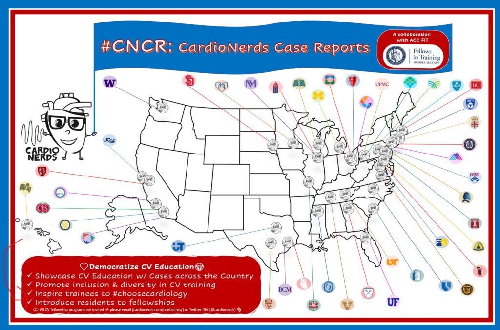
We are teaming up with the ACC FIT Section to use the #CNCR episodes to showcase CV education across the country in the era of virtual recruitment. As part of the recruitment series, each episode features fellows from a given program discussing and teaching about an interesting case as well as sharing what makes their hearts flutter about their fellowship training. The case discussion is followed by both an E-CPR segment and a message from the program director.
CardioNerds Case Reports: Recruitment Edition Series Production Team
-

Bibin Varghese, MD -

Rick Ferraro, MD -

Tommy Das, MD -

Eunice Dugan, MD -

Evelyn Song, MD -

Colin Blumenthal, MD -

Karan Desai, MD -

Amit Goyal, MD -

Daniel Ambinder, MD


The AI-powered Podcast Player

